Pyrite EDS spectrum, 50 kV Rh anode x-ray beam, CSIRO 2015
Loading...
Measurement details
| Description | EDS spectrum of Pyrite |
| Reference | Spectra from unpublished supplementary material for: Torpy A., Wilson N.C., MacRae C.M., Barnes S., and Verrall M. (2015), Characterising drill core mineralogy by micro-XRD mapping, AMAS XIII: the 13th Biennial Australian Microbeam Analysis Symposium: program and abstracts, Eds: Karsten Goemann, Leonid Danyushevsky and Jay Thompson, University of Tasmania, Hobart, February 2015. IBSN: 978-09580408-5-3. https://nla.gov.au/nla.obj-2477868828/view |
| Date measured | 2015-02-04 |
| Material | Pyrite [ FeS2 ] |
| Analysis technique | Energy dispersive x-ray spectrometry (EDS) |
| Instrument | Bruker M4 Tornado |
| Probe | 50 kV Rh anode x-ray beam |
| Anode current | 0.499 mA |
| Detector name | Bruker XFLASH |
| Det. technology | Silicon drift detector (SDD) |
| Real time | 16.591 s |
| Live time | 5.65825 s |
| Dead time | 65.8957 % |
| Channel count | 4096 |
| Energy range | -954.595 … 39979.5 eV |
| Gain | 9.99612 eV/channel |
| Intensity range | 9.162×10⁴ counts |
| Integral | 2.4457×10⁶ counts |
Specimen details
Specimen image
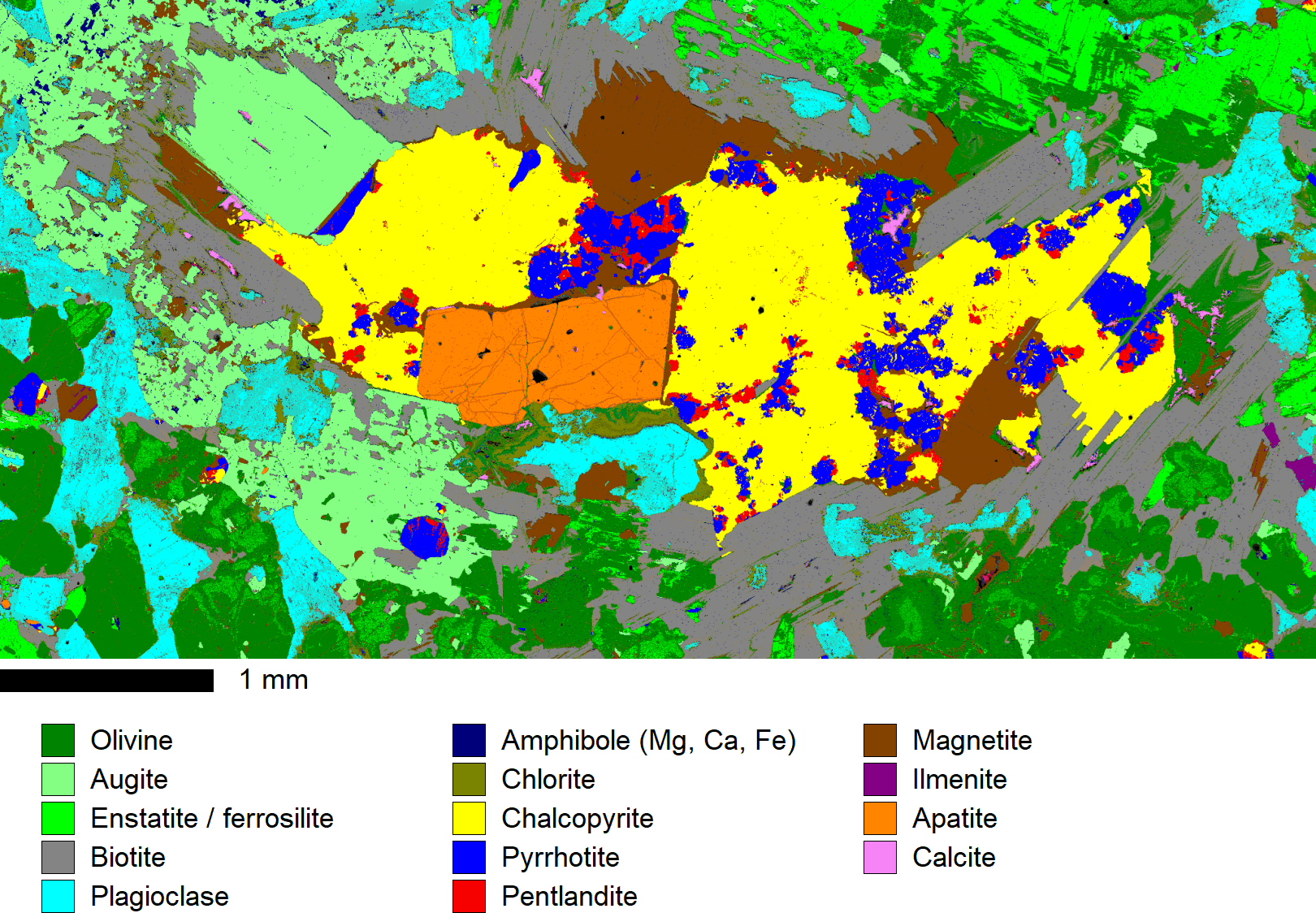
Clustered EPMA phase map of the KH-2 specimen (owner: S. Barnes, CSIRO Mineral Resources), from the Kharelakh intrusion, Noril'sk, Russia
(C) 2015 CSIRO Process Science and Engineering. All Rights reserved.
Composition
| Element | Atoms | At% | Wt% |
|---|---|---|---|
| Fe | 1 | 33.3 | 46.5 |
| S | 2 | 66.7 | 53.5 |
| Total | 3 | 100 | 100 |